Late diagnosis of eye diseases remains one of the leading causes of preventable vision loss worldwide. Many serious conditions such as diabetic retinopathy, glaucoma, and macular degeneration develop gradually and without noticeable symptoms in their early stages. As a result, patients often seek medical attention only after significant damage has already occurred.
Preventing late diagnosis requires advanced tools that can detect subtle changes within the eye before symptoms appear. Retinal imaging has become a critical component of modern ophthalmology, enabling clinicians to identify early signs of disease and intervene at the right time.
The Problem of Late Diagnosis in Eye Care
One of the biggest challenges in eye care is that many diseases are asymptomatic in their initial stages. Patients may have normal vision while structural damage is already progressing in the retina or optic nerve.
For example, diabetic retinopathy begins with microvascular changes that are not visible to the patient. Similarly, glaucoma can cause optic nerve damage long before any noticeable loss of vision occurs. These conditions are often diagnosed late because early warning signs go undetected.
Delayed diagnosis can lead to irreversible damage, making treatment less effective and increasing the risk of permanent vision loss. This highlights the importance of proactive screening and early detection.
Role of Retinal Imaging in Early Detection
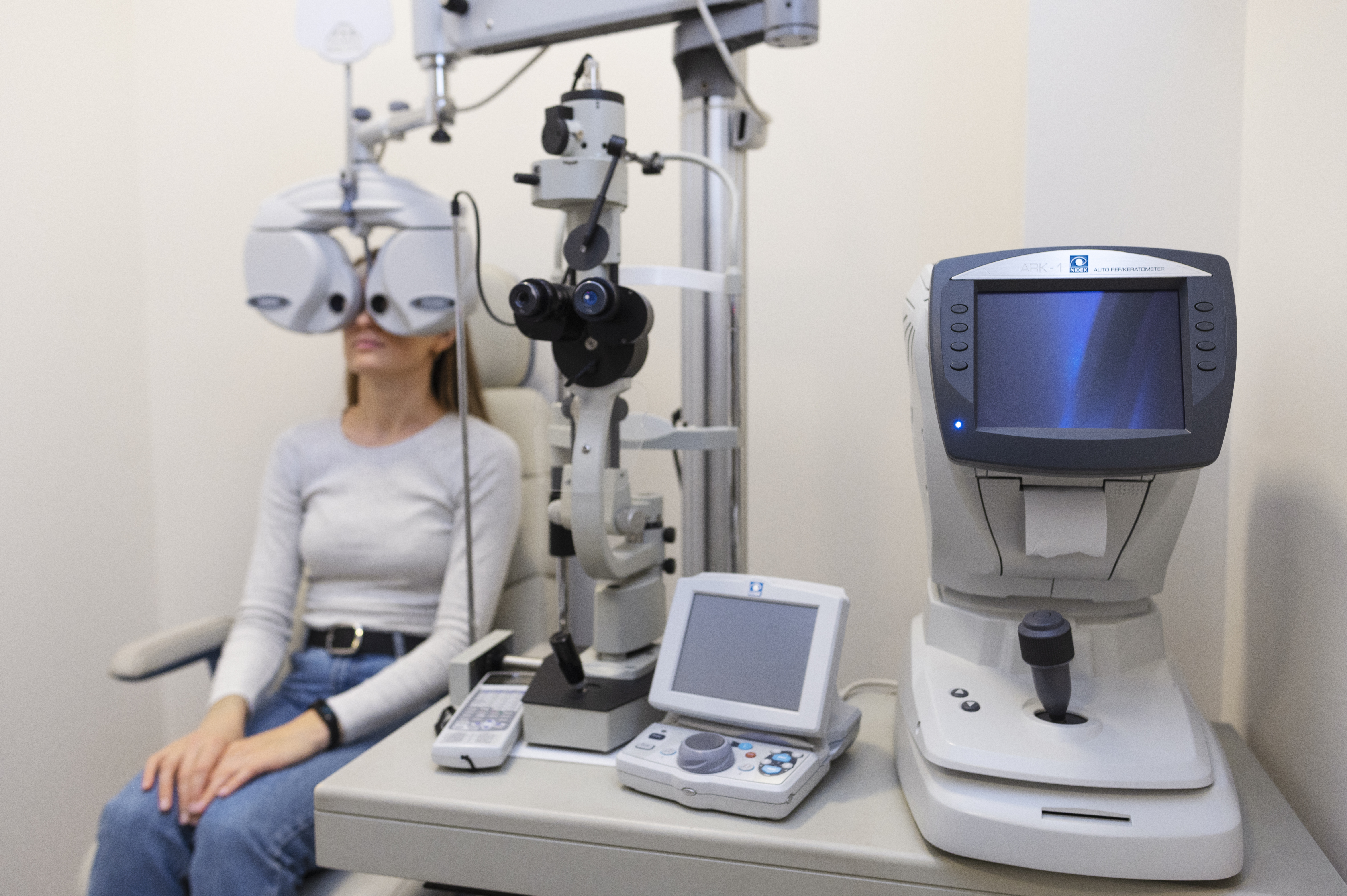
Retinal imaging provides a detailed view of the internal structures of the eye, including the retina, optic nerve, and blood vessels. This allows clinicians to detect abnormalities that may not be visible during routine eye examinations.
Fundus photography is widely used for documenting the condition of the retina. It captures high-resolution images that serve as a baseline for comparison over time. These images help identify subtle changes and track disease progression.
By enabling early detection, retinal imaging plays a crucial role in preventing late diagnosis and improving patient outcomes.
Importance of Advanced Imaging in Preventing Delayed Diagnosis
A Fundus Photography Camera is a key diagnostic tool that captures detailed images of the retina, allowing clinicians to identify early signs of eye diseases with high accuracy. These images provide a comprehensive view of retinal structures, making it easier to detect abnormalities at an early stage.
Through this imaging technology, specialists can identify:
- 1. Microaneurysms and early vascular changes
- 2. Subtle optic nerve damage
- 3 .Initial stages of macular degeneration
- 4. Small hemorrhages or lesions
- 5. Early signs of retinal swelling
These findings are often present long before symptoms develop. Detecting them early allows for timely intervention, reducing the risk of late diagnosis.
Supporting Proactive Screening Programs
One of the most effective ways to prevent late diagnosis is through regular screening programs. Retinal imaging plays a vital role in these programs by providing a quick and reliable method for examining the retina.
Fundus imaging is non-invasive and efficient, making it suitable for large-scale screenings. It allows clinicians to identify at-risk individuals and recommend further evaluation or treatment as needed.
Screening programs using retinal imaging have been shown to significantly reduce the incidence of advanced eye diseases by promoting early detection and intervention.
Monitoring Disease Progression
Preventing late diagnosis is not only about detecting disease early but also about monitoring its progression. Chronic eye conditions require continuous observation to ensure that they do not worsen over time.
Fundus imaging provides a reliable way to track changes in the retina. By comparing images from different visits, clinicians can assess:
- 1. Progression of disease
- 2. Development of new abnormalities
- 3. Effectiveness of treatment
- 4. Stability of existing conditions
This ongoing monitoring helps ensure that any changes are identified promptly and addressed before they lead to severe complications.
Enhancing Diagnostic Accuracy
Traditional eye examinations can sometimes miss subtle changes, especially in the early stages of disease. Fundus imaging enhances diagnostic accuracy by providing clear and detailed visual data.
These images can be reviewed by multiple specialists, reducing the risk of misdiagnosis. Additionally, digital imaging allows for better documentation and analysis, improving the overall quality of care.
Artificial intelligence is also being integrated into retinal imaging systems, enabling automated detection of abnormalities and supporting clinicians in making more accurate diagnoses.
Improving Patient Awareness and Compliance
Visual evidence plays a significant role in patient education. When patients can see images of their own retina, they are more likely to understand their condition and the importance of treatment.
Fundus imaging helps improve patient awareness by providing clear visual representation of eye health. This encourages patients to follow medical advice, attend regular check-ups, and adhere to treatment plans.
Improved patient compliance is essential for preventing disease progression and avoiding late diagnosis.
Why Reliable Imaging Solutions Matter
The effectiveness of preventing late diagnosis depends on the quality of imaging equipment. High-resolution images are essential for detecting subtle changes and ensuring accurate diagnosis.
Matronix Optotechnik provides advanced ophthalmic diagnostic solutions designed to support early detection and efficient monitoring of eye diseases. Their imaging systems deliver precise and detailed retinal images, enabling clinicians to diagnose conditions at an early stage and prevent complications. With a focus on innovation and reliability, they contribute to improved patient outcomes and better clinical workflows.
Conclusion
Late diagnosis of eye diseases can have serious consequences, often leading to irreversible vision loss. Since many conditions develop silently, early detection is essential for effective management.
Retinal imaging plays a crucial role in preventing delayed diagnosis by providing detailed insights into the health of the retina. It enables clinicians to identify early signs of disease, monitor progression, and guide timely treatment.
As technology continues to advance, the role of imaging in eye care will become even more significant. Regular screenings and access to high-quality diagnostic tools remain key to preventing vision loss and ensuring better eye health outcomes.
Powered by Froala Editor
You may also like
More from this category.

Common Problems in Sewage Treatment Plants and Their Solutions

Best ENT Specialist in Lahore Near Me | Dr. Zahra Aleem

How Best Dentists in California Build Long-Term Patient Trust

Conquering the Infinite Descent: A Guide to the Addictive World of Slope Game

Legal DNA Test for Paternity,Immigration & Family Law

Best Areas to Buy Property in Dubai: Complete Area Guide (2026)

Best Skin Clinic in Jubilee Hills – Expert Care for Every Skin Type

Wellness Coach Certification: Why Becoming a Wellness Coach is a Future-Ready Career Choice

Natural Lip Injections: A Guide to Natural-Looking Results